NUCLEAR MEDICINE

WHAT IS NUCLEAR MEDICINE?
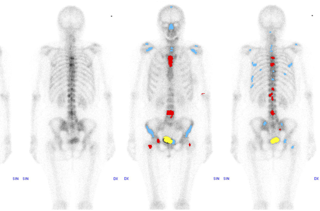
Nuclear medicine uses a small amount of a radioactive substance to produce images of the body. The radioactive material is detected in the body by the scanner and then images mapping the material throughout the body are produced. These images are two- or three-dimensional and show detailed body anatomy and function. Nuclear scans allow clearer images of organ and tissue structure than traditional, non-contrast imaging.
What is nuclear medicine used for?
Nuclear scans can be used to evaluate and diagnose a variety of conditions and diseases. Some of these include:
- Tumors and infection
- Bone disease and fractures
- Thyroid disease
- Respiratory and blood-flow problems in the lungs
- Functional studies of the kidney, bowel, gallbladder and heart
How to prepare for a nuclear medicine study
Because nuclear medicine requires an injection of a small dose of radioactive material, some special preparation is required before your scan. To prepare for a nuclear medicine study, please:
- Bring a copy of the order for the procedure from your referring physician, your insurance card, and photo identification.
- On the day of your exam, wear comfortable, loose-fitting clothing.
- You should drink plenty of water before the test.
- Take your usual medications.
- If the examination is done to evaluate the stomach or gallbladder, do not eat or drink for 4 hours before the test.
- Always inform your doctor and technologist if you are pregnant or could be pregnant.
What to expect during a nuclear medicine study
Typically, a nuclear medicine exam takes about 20-45 minutes; however, some specialized studies may last several days. A radiopharmaceutical, known as a tracer, is usually administered either intravenously or by mouth. Although usually done with a small needle, some patients experience minor discomfort from the intravenous injection. Most of the radioactivity is expelled out of your body in urine or stool. The rest simply disappears over time.
For most nuclear scans, you will lie down on a table, and a nuclear imaging camera will be used to capture the image of the area being examined. The camera is either suspended over or below the exam table or in a large donut-shaped machine, similar to a CT scanner. During the scan, you must remain as still as possible. You will hear low-level clicking or buzzing noises while you are in the machine.
When your examination is over, you may resume your normal daily activities unless otherwise instructed by your doctor. One of our board-certified radiologists will review the images and send a report to your doctor.
What if I’m claustrophobic?
Some claustrophobic patients may experience a “closed in” feeling during a scan. If this is a concern, please let us know prior to your appointment. Conscious sedation may be available, depending on your preferred location.